

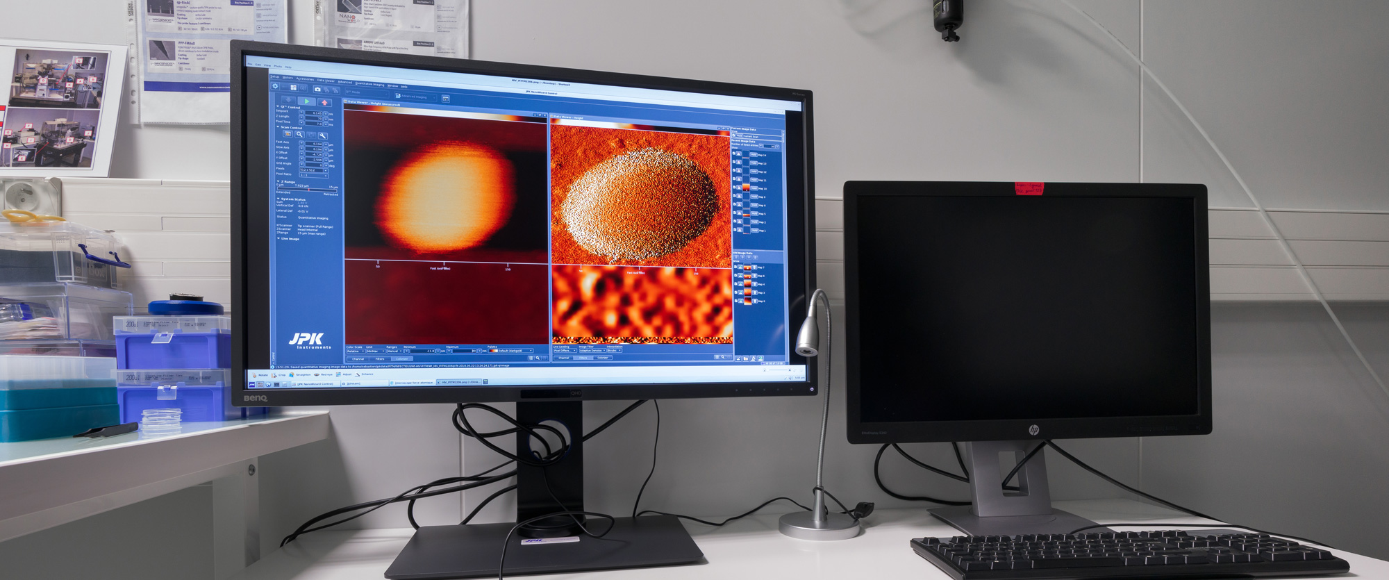
AFM / Optical Microscope (BIO-AFM) for L3 imaging of infected living cells and class 3 pathogens (viruses and bacteria).
Possible samples:
fixed or living cells in their culture medium, bacteria, yeasts, macromolecules (nucleic acids, proteins), polymers, fibers, composites, lipid films, lipid vesicles (GUV, LUV), extracellular vesicles (exosomes), structures purified biologics (viral capsids for example), virus-like-particles (VLPs), class 2 and 3 viruses.
Size of the smallest objects detectable on the surface: a few nanometers X and Y
Height of observable objects: from 0.5nm to 12ÎĽm
Dimension of square images: from 50nm to 100ÎĽm
Sizes of the observable samples: 30mm x 50mm in X and Y, 5 to 10mm in Z
Possibility of working in liquid or air in a thermostatic chamber.
AFM images :
a) Chikungunya virus
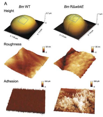
b) Bacteria
c) HEK293T cell
Publications:
Transportin-1 binds to the HIV-1 capsid via a nuclear localization signal and triggers uncoating.
Fernandez J, Machado AK, Lyonnais S, Chamontin C, Gärtner K, Léger T, Henriquet C, Garcia C, Portilho DM, Pugnière M, Chaloin L, Muriaux D, Yamauchi Y, Blaise M, Nisole S, Arhel NJ.
Nat Microbiol. 2019 Oct 14. doi: 10.1038/s41564-019-0575-6.
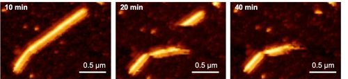
Figure: In vitro reconstituted HIV-1 proteins using recombinant capsid were contacted with purified Transportin. The images taken by Atomic Force Microscopy illustrate the deformations and breaks by HIV-1 capsid representing (left to right) 10, 20 and 40 min after injection.
Safia Ouahrani-Bettache, MarĂa P. JimĂ©nez De BagĂĽĂ©s, Jorge De La Garza,Luca Freddi, Juan P. Bueso, SĂ©bastien Lyonnais, Sascha Al Dahouk, Daniela De Biase, StephanKöhler & Alessandra Occhialini. 2019 , Virulence, 10:1, 868-878, DOI:10.1080/21505594.2019.1682762
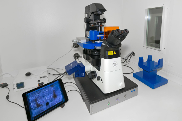
Optical Microscope NIKON Ti2-U inverted Microscope with Phase Contrast and Fluorescence
- Nikon CFI Lens Achromat LWD ADL-20x Ph1-
- Nikon CFI Achromat LWD ADL-40x Ph2
- NIKON CFI APO VC 100x oil (A.N. 1.4 DT 0.13)
White LED light source and Epi-Fluorescence Intensilight 100w (Hg, manual)
- Cubes Filters:o GFP (Ex466 / 40, DM 495, stop filter BA 525/50)
- mCHERRY, mRFP (Ex562 / 40, DM 593, BA 640/75 stop filter)
- CY5, APC, DiD, Alexa Fluor 647, Alexa fluor 660 (Ex628 / 40, DM 660, BA 692/40 stop filter)
JENOPTIK Fluorescence ProgRes MFcool Camera (2/3 “1.4 MegaPixel CCD, 1350×1024 pixel resolution, 14bit)
 Chargement en cours ...